Overview of ServicesThe Translational Bioimaging Resource (TBIR) is housed in the basement of the Biosciences Research Labs (BSRL) building. The 12,000 sq. ft. TBIR serves as a university-wide resource for pre-clinical biomedical imaging. The resource has the capability to image small biological constructs, small and large animals, and humans. For imaging small biological constructs and small animals the TBIR includes intra vital microscopy, MRI, ultrasound, CT, PET, SPECT, and bioluminescence. For imaging larger animals and humans the resource includes MRI and ultrasound. The resource includes personnel to help you with project development and to help you use the equipment. |
|
|
|
Human MRIThe TBIR is dedicated to supporting MRI research within the University community and for industry partners in pharmaceuticals, health care, veterinary care, and behavioral sciences. The TBIR houses a 3 Tesla Siemens Skyra VE11 that is able to support work that addresses a wide range of research questions, including questions related to arthritis, traumatic brain injuries, PTSD, cancer treatment efficacy, depression, kidney transplant viability, CPR, Alzheimer’s, complex grief, and healthy aging. |
|
|
Small Animal MRIThe TBIR manages a 7T Bruker Biospec research MRI scanner. Users can perform physiological and functional imaging of soft tissue anatomy, tumor growth, angiogenesis, cellularity, inflammation, myelination, myocardial function, early therapeutic effects, extracellular pH, redox state, and other applications. 3D anatomical imaging of soft tissues is performed at up to 300 μm resolution. The facility has technical support to aid in surgical procedures and anesthesia, protocol modifications and compliance, safety training, and consultations. A Cubresa NuPET MRI compatible PET insert enables evaluation of tumor therapy response through diffusion-weighted MRI with PET tracers specific for tumor proliferation. The PET insert can also improve tumor detection through diffusion-weighted and T2-weighted MRI with PET tracers for specific biomarkers as well as evaluate drug delivery and perfusion for molecular theranostics assessments. |
|
|
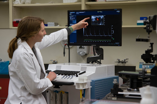
High Resolution UltrasoundThe TBIR manages a VisualSonics Vevo 3100 along with data management and analysis software. This small-animal ultrasound device provides axial resolution down to 30 microns. The system can be used to measure a variety of cancer-related metrics such as tumor growth, tumor blood flow and volume, angiogenesis, and molecular imaging with microbubble contrast agents. Cardiovascular imaging and measurement of cardiac function are two other popular applications. The technology also applies to other disciplines including neurosciences, embryology, and ophthalmology. Specialized human studies are also possible with institutional review board approval. |
|
|
MicroCTThrough a partnership with the University of Arizona Cancer Center, the TBIR provides a Siemens Inveon micro-CT scanner, which is a variable zoom cone-beam X-ray CT system that can generate images with a spatial resolution as small as 20 microns over a field of view as large as 8.4 cm x 5.5 cm. The large field of view enables single-bed position imaging of an entire mouse as well as larger objects such as rats with bed-stitching. The fully shielded design has built-in anesthesia and optional physiological monitoring. The scanner is best at evaluating bone density, but it can also image soft tissues with the use of CT contrast agents. Physiological monitoring ensures viability of the animal during the scan, and respiratory & cardiac gating improves image quality of the heart and lungs. |
|
|
Bioluminescence and Fluorescence ImagingThrough a partnership with the University of Arizona Cancer Center, the TBIR provides a Spectral Instruments Imaging Lago, which has powerful multi-modality imaging capabilities in an advanced, user-friendly system. The turn-key system offers bioluminescence and fluorescence imaging. The high performance 2048x2048 air-cooled CCD camera provides high sensitivity with low background noise for accurate quantitative analysis. The Lago has 14 excitation wavelengths ranging from 360-805 nm and 20 emission filters ranging from 490-870 nm. The system has a field-of-view ranging from 6x6 cm up to 25x25 cm. These dynamic filter combinations can support a wide range of applications ranging from well-plate imaging to small animal imaging. Bioluminescence imaging of reporter genes such as luciferase and green fluorescent protein are easily accomplished with this scanner, and imaging of exogenous fluorescent agents can also be performed. The Lago incorporates a heated imaging platform and built-in isoflurane gas anesthesia. |
|
|
Quantitative Digital HistologyThrough a partnership with the University of Arizona Cancer Center the TBIR offers access to Definiens Tissue Studio 4.2 for biomarker and morphological profiling in histological images. Tissue Studio is an easy to use, robust, and powerful tool to automatically detect regions of interest based on user-defined criteria. It can distinguish cells and sub-cellular objects within target regions, and determine morphology and expression profiles per individual cell or cell compartment. It is compatible with brightfield or immunofluorescence images and whole tissue slides or tissue microarrays. Tissue Studio is compatible with most major file formats including Aperio (.svs), Hamamatsu (.ndpi), Leica (.scn), Roche Ventana (.bif), Zeiss (.czi), and TIFF. |
Leadership |
|
|
Andy Rouse, PhD Director BSRL 77 Email: rouse@arizona.edu Phone: (520) 621-4247 |
Scott Squire Clinical MRI BSRL 72 Email: ssquire@arizona.edu Phone: (520) 245-6272 |
Christy Howison Small Animal MRI BSRL 79 Email: chowison@arizona.edu Phone: (520) 626-6653 |
Brenda Baggett BioLum, uCT, & Tissue Studio BSRL 78 Email: baggett@arizona.edu Phone: (520) 626-5052 |
| Hours | Location |
|
Monday - Friday 9:00am - 5:00pm 24/7 access for trained users |
Bioscience Research Laboratory, Basement 1230 N Cherry Ave. Tucson, AZ 85719 |
| Name | Role | Phone | Location | |
|---|---|---|---|---|
| Translational Bioimaging Resource |
General Facility Contact
|
520-621-4247
|
rouse@arizona.edu
|
BSRL Room 77
|
| Core Facility Business Office |
Billing Inquiries
|
520-621-4064
|
CoreBusiness@arizona.edu
|
Keating Room 200
|
| Service list |
| ► Clinical MRI (1) | |||
| Name | Description | Price | |
|---|---|---|---|
| Human MRI Contrast Study |
Add-on charge to cover materials used to scan a single MRI particpant using contrast. |
||
| ► Bioluminescence and Fluorescence Imaging (1) | |||||||||||||||||||||
|
|||||||||||||||||||||
| ► Clinical MRI (3) | |||||||||||||||||||||
|
|||||||||||||||||||||
| ► Confocal & Multiphoton Microscopy (1) | |||||||||||||||||||||
|
|||||||||||||||||||||
| ► High Resolution Ultrasound (1) | |||||||||||||||||||||
|
|||||||||||||||||||||
| ► Quantitative Digital Histology (1) | |||||||||||||||||||||
|
|||||||||||||||||||||
| ► Small Animal MRI (1) | |||||||||||||||||||||
|
|||||||||||||||||||||
| ► Small Animal MicroCT (2) | |||||||||||||||||||||
|
|||||||||||||||||||||